Welcome to the Science Core Facility!

We are located in the Science Center, Harned Hall, which opened in May 2006 and are home to the Weatherwax Electron Microscopy Suite. The Science Core Facility provides outstanding facilities and equipment for both teaching and research for the entire campus.

Some of our resources include:

  • Hitachi S3400N Variable Pressure Scanning Electron Microscope
  • Markforged The Mark Two 3D Printer
  • Nikon D-Eclipse C1 Confocal microscope
  • BioRad CFX96 Real-Time PCR Detection System
  • LI-COR Odyssey Fc Dual Mode Imaging System
  • GenePix 4100A Microarray Scanner
  • SpectraMax M2 Microplate Reader
  • Bio-Rad GelDoc XR+

The science core facility is a campus-wide resource for students, faculty and staff. The core is also open to academic and commercial users outside of the University of Puget Sound. Outside users need to contact the Science Core Facility Technician directly for training and appointments. For more information on some of our instruments please see our Equipment page.
Student Images

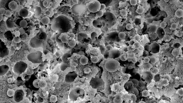
Scanning Electron Micrograph of eggshell surface - People's Choice 2019

Smooth spheres found on S. virgatus eggshell surface at 0 weeks of incubation by Grace Elliott '20. The spheres vary in shape and size and are often located in the troughs that lie between the raised polygons of the shell surface. 

Scanning Electron Micrograph of Wolframite - People's Choice 2016

A rock of unknown composition was identified as the mineral wolframite (FeWO4) by Miriam Cohen '19 using energy dispersive spectroscopy on our Hitachi S3400N SEM. The needlelike features in the center is uncommon for mineral growth and an interesting find.

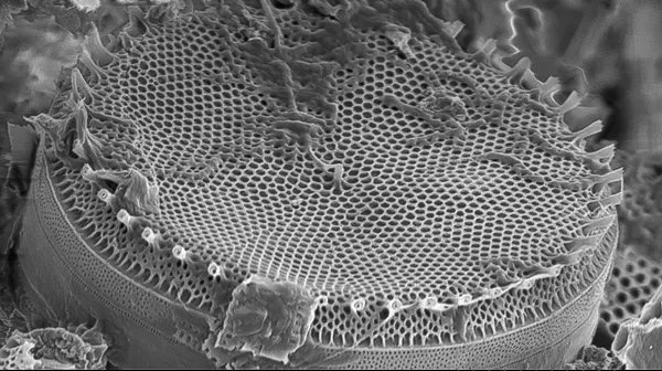
Scanning Electron Micrograph of a Diatom - People's Choice 2015

This diatom was found in a sediment sample by Blake Hessel '16 and imaged on our Hitachi S3400N SEM. It is fascinating to observe the fine detail of the various surface patterns and embellishments.

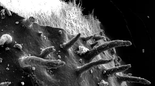
Scanning Electron Micrograph of Pedicellaria – Judge's Choice 2014

P. ochraceous pedicellaria responding to contact with another species of sea star taken by Haila Schultz ’16 on our Hitachi S3400N SEM. These comparisons will shed light on competition between sea star species and development of pedicellaria size.

Scanning Electron Microscopy of Lichen Holdfasts - Winner Fall 2013

Observation of tissue segregation in a folios lichen (Parmelia spp.) taken by Kelsey Crutchfield-Peters '14 on our Hitachi S3400N SEM. It is important in studying how tissue structure, and symbiont abundance, changes within the lichen in response to environmental factors. 

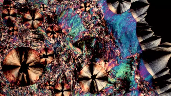
Polarizing Light Microscopy of Liquid Crystals – Winner Fall 2013

Liquid crystal compound synthesized by Mary Packard ’15 being cooled from the nematic phase, which is one of the liquid crystal phases,  into regular crystals. Image was taken on a Nikon Labophot-2-Pol  Polarizing Transmitted Light Microscope we have on campus.